
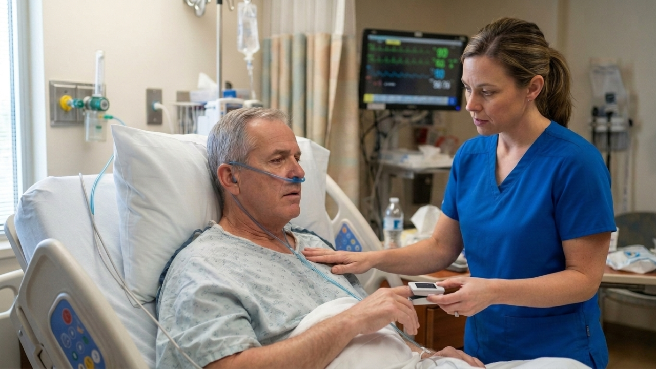
The monitor reads SpO2 86 percent, the arterial blood gas is altered, and you freeze on which label to write: impaired gas exchange, ineffective airway clearance, or ineffective breathing pattern. That moment is where most students get stuck, and the BP-style shortcut of matching the disease to the diagnosis does not work here.
Keep one distinction front and center. Impaired gas exchange is a nursing diagnosis, not a medical diagnosis. It describes a problem at the alveolar-capillary membrane, the place where oxygen and carbon dioxide cross between the lungs and the blood. It is not the disease behind it, whether that is COPD, pneumonia, a pulmonary embolism, heart failure, or ARDS. This guide walks you through how to confirm the gas-exchange membrane is the problem, how to read the gas, how to tell the three respiratory diagnoses apart, and how to build a care plan with goals you can actually measure.
Bottom line: Impaired gas exchange is a problem of oxygenation and carbon dioxide at the cellular level, shown by an abnormal ABG or SpO2, not just by a lung disease on the chart. Distinguish it from ineffective airway clearance (secretions) and ineffective breathing pattern (mechanics), prioritize airway and oxygenation first, and titrate oxygen carefully in patients who retain carbon dioxide.
In This Article:
- What impaired gas exchange means in a care plan
- The three respiratory diagnoses, side by side
- Nursing diagnosis vs. medical diagnosis
- Assessment data to collect first
- How to read the ABG
- How to choose the right diagnosis
- Priority and related nursing diagnoses
- How to prioritize
- A worked patient scenario
- Sample care plan table and interventions
- Patient education, common mistakes, and example PES statements
What Impaired Gas Exchange Means in a Nursing Care Plan
Impaired gas exchange is an excess or deficit in oxygenation or carbon dioxide elimination at the alveolar-capillary membrane.
In plain language, gas exchange works only when ventilation and perfusion match. Ventilation is air reaching the alveoli. Perfusion is blood reaching the pulmonary capillaries. When the two do not line up, oxygen and carbon dioxide cannot move efficiently across the membrane. That mismatch is the heart of the problem.
There are two ways the match breaks down. Dead space is ventilation without perfusion: the alveoli fill with air, but no blood arrives to pick up the oxygen, as in a pulmonary embolism. Shunt is the reverse, perfusion without ventilation: blood flows past alveoli that are not being ventilated, as in pneumonia, atelectasis, or a mucus plug. Both leave the patient hypoxemic, and the cause points you toward different interventions.
Two more terms get mixed up constantly. Hypoxemia is low oxygen in the blood, an ABG or SpO2 finding; think of it as a localized issue. Hypoxia is low oxygen at the tissue level, often seen with decreased kidney function or a change in mental status; think of it as a systemic problem. Impaired gas exchange usually starts as hypoxemia and can progress to hypoxia if it is not corrected. The initial interventions are the same: restore oxygenation.
Download our Normal vs. Shunt vs. Dead Space Diagram.
Important: Central cyanosis (a blue tongue and oral mucosa), a falling level of consciousness, an SpO2 that drops fast and does not respond to oxygen, or any sign of respiratory failure is an emergency. Escalate per facility protocol rather than continuing routine care.
Impaired Gas Exchange vs. Ineffective Airway Clearance vs. Ineffective Breathing Pattern
Use your assessment data, not your assumptions, to choose the correct diagnosis. Use one rule for each.
Impaired gas exchange is a problem of oxygenation or carbon dioxide at the alveolar-capillary membrane, and the evidence is an abnormal ABG or SpO2. Ineffective airway clearance is a problem of secretions, and the evidence is retained sputum, adventitious breath sounds, and a weak or wet cough. Ineffective breathing pattern is a problem of the mechanics of breathing, and the evidence is an abnormal rate, depth, or rhythm: tachypnea, bradypnea, accessory muscle use, or an irregular pattern with no obstruction or oxygenation problem.
| Diagnosis | Core problem | Hallmark assessment cue | Example cause | First-line nursing focus |
|---|---|---|---|---|
| Impaired gas exchange | Membrane / diffusion | Abnormal ABG and SpO2 | Pneumonia, ARDS, PE | Oxygenation and positioning |
| Ineffective airway clearance | Secretions / obstruction | Retained sputum, crackles, ineffective cough | Pneumonia, COPD, post-op | Airway clearance and coughing |
| Ineffective breathing pattern | Mechanics of breathing | Abnormal rate, depth, or rhythm; no obstruction or oxygenation problem | Anxiety, pain, neuromuscular | Breathing retraining and positioning |
Note: The same patient often has more than one of these at once. A patient with COPD may have all three. Prioritize the one with the most dangerous evidence, which is usually impaired gas exchange when the ABG or SpO2 is abnormal.
Nursing Diagnosis vs. Medical Diagnosis
The medical diagnosis is the disease: COPD, pneumonia, pulmonary embolism, asthma, ARDS, heart failure, or pulmonary edema. The nursing diagnosis describes the patient’s response to it: impaired gas exchange, ineffective airway clearance, activity intolerance, anxiety, or decreased cardiac output.
Here is the key move. Do not write impaired gas exchange just because the patient has a lung disease. Choose it only when the assessment data support a membrane-level oxygenation or carbon dioxide problem: an abnormal ABG, a low SpO2, cyanosis, or a mental status change from hypoxia.
Three short examples show how the same disease splits:
- A pneumonia patient with an SpO2 of 84 percent and a PaO2 of 55 fits impaired gas exchange.
- A pneumonia patient who is oxygenating fine but cannot clear thick secretions fits ineffective airway clearance.
- An anxious post-op patient with a fast, shallow, splinted breathing pattern but normal gases fits ineffective breathing pattern.
Same chart, three different care plans.
Assessment Data to Collect First

The diagnosis comes after the assessment, not before. Document both what the patient tells you and what you measure.
Subjective cues: reports of shortness of breath or air hunger, fatigue, dizziness, headache (which can signal carbon dioxide retention), feeling anxious or unable to get enough air, and difficulty speaking in full sentences.
Objective cues: the SpO2 trend; ABG values; respiratory rate, depth, effort, and rhythm; accessory muscle use, nasal flaring, or retractions; breath sounds (crackles, wheezes, diminished); skin and mucous membrane color (pallor, central vs. peripheral cyanosis); level of consciousness and mental status; heart rate, rhythm, and blood pressure; capillary refill; and cough and sputum (amount, color, consistency).
Tip: Learn the early and late signs of hypoxia, because students reverse them. Early signs are restlessness, anxiety, tachypnea, tachycardia, mild hypertension, and pale skin. Late signs are confusion or lethargy progressing to loss of consciousness, bradypnea, bradycardia, hypotension, and cyanosis. Cyanosis is a late sign, not an early one.
Tests you may review include pulse oximetry, ABG and VBG, capnography or end-tidal carbon dioxide, chest X-ray, a CBC (hemoglobin matters, because anemia limits oxygen carrying), and the PaO2/FiO2 ratio for ARDS severity.
Nurses do not diagnose from a single finding. They group related cues first. A few clusters that point somewhere specific:
- Low SpO2 plus an abnormal ABG plus crackles plus dyspnea at rest points to impaired gas exchange.
- A productive wet cough plus coarse crackles plus an SpO2 that improves after the patient clears secretions points to ineffective airway clearance.
- Fast, shallow, splinted breathing plus a normal SpO2 and ABG plus recent surgery or anxiety points to ineffective breathing pattern.
How to Read the ABG
Understanding how to interpret arterial blood gases will help you choose the correct nursing diagnosis. Here is a brief review. Start with the normal values.
| ABG parameter | Normal range |
|---|---|
| pH | 7.35 to 7.45 |
| PaCO2 | 35 to 45 mm Hg |
| HCO3 | 22 to 26 mEq/L |
| PaO2 | 80 to 100 mm Hg |
| SaO2 | 95 to 100 percent |
Then work a simple stepwise method. One common memory aid is ROME: Respiratory Opposite, Metabolic Equal. Look at the pH first (acidotic or alkalotic), then the PaCO2 (the respiratory driver), then the HCO3 (the metabolic driver), then decide whether compensation is present, then check the PaO2 and SaO2 for oxygenation.
A few worked examples, labeled as examples only:
- pH 7.30, PaCO2 55, HCO3 24: uncompensated respiratory acidosis with hypoventilation.
- pH 7.36, PaCO2 60, HCO3 34: compensated respiratory acidosis, the classic chronic COPD picture.
- pH 7.32, PaCO2 38, HCO3 18, PaO2 60: metabolic acidosis with hypoxemia.
Tie each one back to the question that matters for your diagnosis: does the gas support a membrane oxygenation problem? A low PaO2 or SaO2 alongside the acid-base picture is what backs impaired gas exchange.
How to Choose the Right Nursing Diagnosis
Work this top to bottom. It is a decision guide, not a substitute for clinical judgment.
- Is the SpO2 or ABG abnormal, pointing to an oxygenation or carbon dioxide problem at the membrane? If yes, impaired gas exchange is supported.
- Are retained secretions or an ineffective cough the main problem? Add or prioritize ineffective airway clearance.
- Is the rate, depth, or rhythm of breathing the main problem, with normal gases? Consider ineffective breathing pattern.
- Are there cardiac signs (edema, abnormal heart sounds, poor perfusion)? Consider decreased cardiac output.
- Is the patient unable to sustain breathing on their own? Consider impaired spontaneous ventilation or the risk version.
Note: A patient often carries several of these at once. The care plan prioritizes the most urgent first, which the prioritization section below works through using ABC logic.
Priority and Related Nursing Diagnoses for Impaired Gas Exchange
These are chosen from the assessment, not by default. Each one below follows the same shape so you can scan it: when it fits, the cues that support it, a sample PES statement, and the student mistake to avoid.
Impaired Gas Exchange (Actual)
The star diagnosis. Use it when the assessment shows an oxygenation or carbon dioxide problem at the membrane: a low SpO2, an abnormal ABG, cyanosis, or a hypoxia-related mental status change. Connect the cause to the mechanism in plain language, for example alveolar fluid in pneumonia creating a shunt. Sample PES statement: “Impaired gas exchange related to alveolar-capillary membrane changes from pneumonia as evidenced by SpO2 86 percent on room air, PaO2 56 mm Hg, crackles, and restlessness.” Common mistake: choosing it just because the patient has a lung disease, with no abnormal ABG or SpO2 to back it.
Ineffective Airway Clearance
Use this when the problem is secretions or obstruction: retained sputum, coarse crackles, and a wet or weak cough. Interventions center on coughing techniques, hydration to thin secretions, suctioning, and chest physiotherapy. Common mistake: labeling it impaired gas exchange when the gases are normal and the real issue is clearing mucus.
Ineffective Breathing Pattern
Use this when the mechanics are abnormal, the rate, depth, or rhythm, from pain, anxiety, neuromuscular weakness, or splinting, with normal gases. Interventions focus on positioning, pursed-lip and diaphragmatic breathing, pain control, and anxiety reduction. Common mistake: this is almost never the primary diagnosis, but new nursing students want to choose it because it is obvious. An altered breathing pattern is present in all patients with a respiratory issue, but this diagnosis is used only when the patient does not have an oxygenation or obstruction problem.
Decreased Cardiac Output
Use this when a cardiac cause drives the hypoxemia, such as heart failure or pulmonary edema. Tie it to perfusion findings (edema, abnormal heart sounds, weak pulses), not to the SpO2 alone.
Activity Intolerance
Use this when the patient desaturates, becomes dyspneic, or fatigues with exertion. Focus on graded activity, monitoring the SpO2 with activity, rest periods, and energy conservation, not generic “exercise more” advice.
Anxiety
Air hunger is frightening, and anxiety worsens dyspnea in a feedback loop. Use this when fear or panic is a clear cue. Interventions: a calm presence, coached breathing, clear explanations, and reducing the work of breathing.
Impaired Spontaneous Ventilation or Risk for Impaired Spontaneous Ventilation
Use this for a patient whose respiratory effort cannot keep up: a rising PaCO2, fatigue, and a declining mental status, who may need noninvasive or mechanical ventilation. This is a high-priority, escalation-focused diagnosis.
Risk for Impaired Gas Exchange
Include one risk diagnosis for the patient who is not yet hypoxemic but has risk factors: immobility, post-op upper-abdominal or thoracic surgery, sedation, or atelectasis risk. A risk diagnosis uses risk factors only and no “as evidenced by.”
How to Prioritize
When a patient carries several diagnoses, order them with the ABCs, safety, and Maslow in mind. Airway and breathing problems sit at the top because they are immediately life-threatening.
- First: airway patency and any signs of respiratory failure or severe hypoxia. Escalate.
- Second: oxygenation and gas exchange. Oxygen therapy, positioning, and treating the cause.
- Third: secretion clearance and breathing mechanics.
- Fourth: activity tolerance, anxiety, and patient education for long-term management.
This is the Next Generation NCLEX clinical-judgment cycle: recognize cues (a low SpO2, an abnormal ABG), analyze and prioritize, take action (oxygen, position, escalate), and evaluate the response. The exam rewards deciding what matters first.
A Worked Patient Scenario
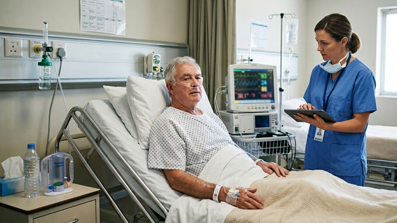
A 68-year-old with community-acquired pneumonia is restless, with a respiratory rate of 28 and an SpO2 of 86 percent on room air, and crackles in the right lower lobe. The ABG shows pH 7.46, PaCO2 32, and PaO2 56.
Walk it through. The cues cluster into a clear oxygenation story (a low SpO2, a low PaO2, restlessness as an early hypoxia sign) with a secretion element (the crackles) and an anxiety element (the restlessness and air hunger). The top candidates are impaired gas exchange, ineffective airway clearance, and anxiety.
Impaired gas exchange leads, because the ABG and SpO2 confirm a membrane oxygenation problem; the low PaCO2 reflects the patient blowing off carbon dioxide as they breathe fast to compensate. A sample SMART goal: the patient will maintain an SpO2 of 92 percent or higher on the ordered oxygen within one hour.
Interventions: apply oxygen and titrate to target, position the patient upright, monitor the SpO2 and respiratory effort, encourage controlled coughing and hydration for the secretions, treat the pneumonia per orders, and use a calm presence to reduce the work of breathing. Evaluate against the SpO2, the respiratory rate, and the patient’s mental status.
Sample Impaired Gas Exchange Nursing Care Plan Table
| Nursing diagnosis | Supporting cues | Short-term goal | Long-term goal | Nursing interventions | Rationale | Evaluation |
|---|---|---|---|---|---|---|
| Impaired gas exchange | Low SpO2, abnormal ABG, restlessness | SpO2 at target on ordered oxygen this shift | Maintains adequate oxygenation without distress | Titrate oxygen, position upright, monitor ABG and SpO2, treat the cause | Upright positioning and titrated oxygen improve V/Q matching | SpO2 and ABG trend toward target |
| Ineffective airway clearance | Retained sputum, coarse crackles, weak cough | Clears secretions with an effective cough | Maintains a patent airway | Hydration, controlled coughing, suctioning as needed | Thinner secretions and an effective cough open the airway | Clearer breath sounds, productive cough |
| Ineffective breathing pattern | Tachypnea, accessory muscle use, splinting | Breathes at an easier rate and depth | Sustains an effective breathing pattern | Positioning, pursed-lip breathing, pain control | Slower controlled breathing reduces air trapping | Respiratory rate and effort improve |
| Activity intolerance | Desaturation and dyspnea with exertion | Completes ADLs without desaturating below target | Tolerates planned activity | Graded activity, SpO2 monitoring with exertion, rest periods | Pacing keeps oxygen demand within supply | Meets activity goal without desaturation |
| Risk for impaired gas exchange | Immobility, post-op atelectasis risk | Maintains a clear airway and target SpO2 | No episode of impaired gas exchange | Incentive spirometry, early mobility, position changes | Lung expansion prevents atelectasis and shunt | SpO2 stays at target; no atelectasis |
Nursing Interventions for Impaired Gas Exchange
Group interventions by purpose instead of repeating them under each diagnosis, and give the reason each one matters.
Monitoring and assessment. Track the SpO2 continuously or frequently, follow the ABG or VBG trend, use capnography where available, and assess respiratory rate, depth, and effort, breath sounds, mental status, and skin color. The reason: trends, not single readings, drive decisions, and a falling mental status is an early hypoxia warning.
Oxygen therapy and delivery devices. Titrate oxygen to the ordered target, commonly an SpO2 of 92 to 96 percent for most adults, or 88 to 92 percent for patients at risk of carbon dioxide retention such as those with COPD. Match the device to the need. Watch for oxygen toxicity with a prolonged high FiO2, generally greater than 50 percent for more than 24 hours.
| Device | Flow rate | Approximate FiO2 | Best use |
|---|---|---|---|
| Nasal cannula | 1 to 6 L/min | About 24 to 44 percent | Stable patients with mild oxygen needs |
| Simple face mask | 5 to 10 L/min | About 40 to 60 percent | Short-term moderate oxygen needs |
| Venturi mask | Device-specific | Precise, about 24 to 50 percent | When an exact FiO2 matters, such as COPD |
| Non-rebreather mask | 10 to 15 L/min | About 60 to 90 percent or higher | Severe hypoxemia and emergencies |
| High-flow nasal cannula / CPAP / BiPAP | High flow or pressure support | Variable, high | Refractory hypoxemia and escalation |
Positioning to improve oxygenation. Use High-Fowler’s or semi-Fowler’s and the tripod or orthopneic position for acute dyspnea, “good lung down” for unilateral lung disease, and prone positioning for ARDS where ordered. The reason: upright and targeted positioning improves V/Q matching and lung expansion.
Airway clearance and breathing techniques. Use pursed-lip and diaphragmatic breathing, incentive spirometry, effective controlled coughing, hydration to thin secretions, suctioning when indicated, and chest physiotherapy. The reason: clearing secretions and recruiting alveoli increases the surface available for gas exchange.
Medications and treating the cause. Bronchodilators, inhaled and systemic corticosteroids, mucolytics, antibiotics for infection, and diuretics for fluid overload, all per orders. The nurse’s role is administration, teaching inhaler technique, and monitoring the response.
Reducing oxygen demand. Cluster care and schedule rest, manage fever and pain (both raise oxygen demand), and address anxiety with a calm presence and coached breathing. The reason: lowering metabolic demand reduces the oxygen the failing system has to supply.
Escalation and emergency readiness. Recognize when oxygen and positioning are not enough, a rising PaCO2, a falling mental status, or refractory hypoxemia, and escalate for noninvasive ventilation or intubation. The reason: early escalation prevents respiratory arrest.
Patient Education Points
- Use prescribed oxygen exactly as ordered, and never increase the flow without contacting your provider, especially with COPD.
- Practice pursed-lip and diaphragmatic breathing, and use the incentive spirometer as taught.
- Pace activities, rest between tasks, and use energy-conservation techniques.
- Stop smoking, and avoid secondhand smoke and air pollutants; ask about cessation support.
- Stay current on influenza and pneumococcal vaccines to prevent respiratory infection.
- Recognize warning signs: worsening shortness of breath, color changes, confusion, or a falling home SpO2, and know when to seek help.
- Take respiratory medications and use inhalers correctly, and bring your devices to appointments for technique checks.
Common Mistakes Nursing Students Make
- Choosing impaired gas exchange just because the patient has a lung disease, without an abnormal ABG or SpO2.
- Confusing impaired gas exchange (membrane and oxygenation) with ineffective airway clearance (secretions) or ineffective breathing pattern (mechanics).
- Confusing hypoxemia (blood) with hypoxia (tissue), and treating cyanosis as an early sign when it is late.
- Cranking up oxygen on a carbon-dioxide-retaining COPD patient instead of titrating to 88 to 92 percent.
- Treating the SpO2 number instead of assessing the whole patient and the trend.
- Writing a vague goal like “patient will breathe normally” instead of a measurable, time-bound outcome.
- Writing interventions without rationales, or listing provider orders without explaining the nurse’s role.
- Adding “as evidenced by” to a risk diagnosis; risk diagnoses use risk factors only.
- Forgetting to reassess and evaluate whether the interventions actually improved oxygenation.
Example PES Statements
These are examples only. Every statement has to match your own patient’s assessment data.
- Impaired gas exchange related to alveolar-capillary membrane changes from pneumonia as evidenced by SpO2 86 percent on room air, PaO2 56 mm Hg, crackles, and restlessness.
- Impaired gas exchange related to ventilation-perfusion mismatch from pulmonary embolism as evidenced by sudden dyspnea, hypoxemia despite supplemental oxygen, and tachypnea.
- Impaired gas exchange related to reduced alveolar surface area from emphysema as evidenced by prolonged expiration, a barrel chest, and an SpO2 of 88 percent.
- Impaired gas exchange related to pulmonary fluid accumulation from heart failure as evidenced by bibasilar crackles, dyspnea at rest, and an ABG showing hypoxemia.
- Risk for impaired gas exchange related to immobility and post-operative atelectasis (risk diagnosis: risk factors only, no “as evidenced by”).
Free Impaired Gas Exchange Care Plan Worksheet
You can practice the full reasoning chain with our impaired gas exchange care plan worksheet, which walks you from cues and the gas to a priority diagnosis and a measurable goal. Download the free Impaired Gas Exchange Care Plan Worksheet here.
Key Takeaways
- Impaired gas exchange is a nursing diagnosis describing an oxygenation or carbon dioxide problem at the alveolar-capillary membrane, not the medical disease.
- Choose it from assessment data, an abnormal ABG and SpO2, not just because the patient has a lung disease.
- Distinguish it from ineffective airway clearance (secretions) and ineffective breathing pattern (mechanics).
- Know your ABG basics and the early-vs-late signs of hypoxia.
- Prioritize airway and oxygenation first, and titrate oxygen carefully in COPD.
- Strong care plans use measurable goals, interventions with rationales, and evaluation.
Patients with COPD are a classic case where all three respiratory diagnoses can collide, and the oxygen question gets its own set of rules. Our nursing diagnosis for COPD guide works through that prioritization in detail.
If you want to think like a nurse under exam pressure, working practice questions is one of the most reliable ways to get there. Our NCLEX-RN practice tests give you realistic practice so the reasoning becomes second nature before test day. All Healthcare Careers will give you the most realistic exam experience possible.